
Advanced Imagery
X-Ray:
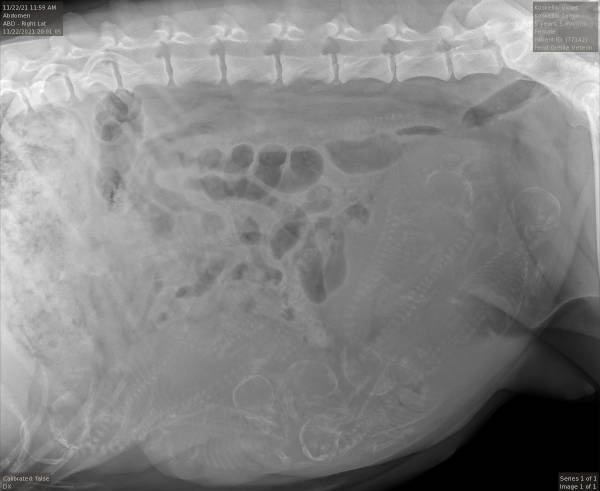
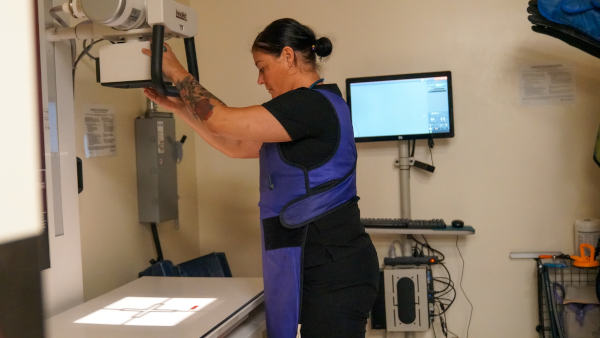
Pend Oreille Veterinary Service is able to offer digital radiology for both dental and body x-rays. This allows for quick imaging in high resolution while minimizing the risks of radiation. X-rays are a non-
invasive diagnostic tool which provides images of multiple parts of the body. We are also able to email these images to specialists when indicated.
Ultrasound:
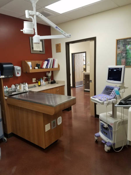
At Pend Oreille Veterinary Service, we are proud to offer ultrasound to help diagnose and manage multiple conditions in our small animal patients.
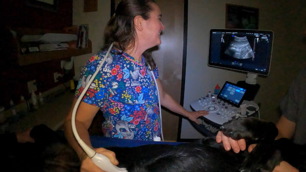
Ultrasound is an imaging technique that uses sound waves that “echo” off of internal structures to create pictures of the body’s soft tissues. Ultrasound is complementary to, but different from, other imaging techniques such as x-ray, CT scan and MRI. Ultrasound is non-painful, non-invasive and considered very safe. Most ultrasound studies can be completed in an awake, cooperative patient. Some pets may require sedation for a complete exam to facilitate them holding still. When biopsies or samples of tissues are taken using ultrasound, general anesthesia may be necessary.
To perform ultrasound, most pets will need to have a small amount of hair clipped from their abdomen or chest. They will need to lay very still on their back or side for the exam. For abdominal ultrasound, fasting is very important- usually 4 hours for cats or 8 hours for dogs.
Ultrasound may be recommended to evaluate for many conditions including:
- Pregnancy
- Urogenital problems
- Trauma evaluation for internal bleeding
- Unexplained weight loss
- Abnormal blood work
- Liver disease
- Adrenal disease
- Vomiting, diarrhea
- Abdominal mass felt on physical exam
- Abnormal xrays
- Fluid in abdomen
- Heart murmur/evaluation of heart disease
Endoscopy:
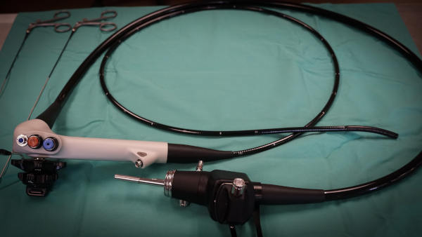
Endoscopy is a non-invasive way to see the internal structure of the tunnel-like systems of the body including the esophagus, stomach, duodenum, colon, nasal passages, pharynx, larynx, trachea and bronchi and ear canals using a high quality camera.
All forms of endoscopy require general anesthesia, so pets are admitted in the morning and spend the whole day with us. They will have an IV catheter placed to administer medications and an endotracheal tube is used to protect the airway while providing gas anesthesia. Careful anesthetic monitoring is provided at all times for endoscopy patients. Endoscopic exams are considered less invasive than general surgery and thus are non to minimally painful. The endoscope can be used to identify and retrieve foreign objects in the nasal passages or respiratory system, as well as the esophagus and stomach. Endoscopy can also be used to evaluate for chronic vomiting and diarrhea by allowing visualization of the lining of GI tract and giving us the ability to collect biopsy samples. Chronic nasal discharge, sneezing, and coughing can also be aided by examining the respiratory system and taking samples for cytology and bacterial culture. Chronic ear conditions can be managed using video otoscopy for deep cleaning, sample collection and evaluation of eardrum and middle ear.
Most pets recover very quickly from endoscopy. For GI patients, mild nausea, bloating and gas may be seen. For respiratory patients, mild nosebleed, congestion or increased cough may be seen temporarily. The doctor will discuss visual findings the day of the procedure. If samples were collected for biopsy, cytology or culture, which will be reported once they are available from the laboratory, often in 7-10 days.